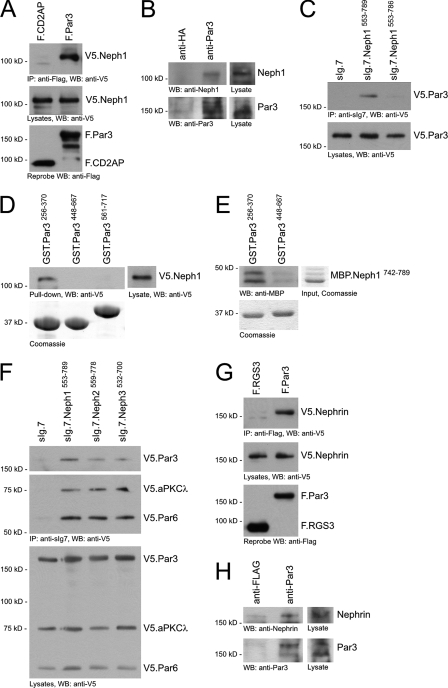
FIGURE 2.
Neph-Nephrin proteins bind to the Par3, Par6, and aPKC complex. A, V5.Neph1 and FLAG-tagged full-length CD2AP or Par3 were expressed in HEK 293T cells. After immunoprecipitation with anti-FLAG antibody, the immobilized Neph1 was detected with anti-V5 antibody in the precipitate containing Par3 but not CD2AP. WB, Western blot. B, lysate from mouse glomeruli was immunoprecipitated with a control polyclonal antibody (rabbit anti-hemagglutinin (anti-HA)) or rabbit anti-Par3 antibody, respectively. Immobilized Neph1 was detected with a Neph1-specific antiserum. C, deletion of the last three amino acids of Neph1, the PDZ domain-binding motif, abrogated binding of Par3 to the cytoplasmic Neph1 tail. D, lysate of HEK 293T cells transfected with V5.Neph1 cDNA was subjected to a pull-down assay with recombinant affinity-purified PDZ domains of Par3 fused to GST. Neph1 specifically interacted with the first PDZ domain of Par3 (Par3256–370). The lower panel shows expression levels of GST fusion proteins on a Coomassie Blue-stained gel. E, this interaction was confirmed by in vitro interaction assay with recombinant affinity-purified carboxyl terminus of Neph1 (MBP.Neph1742–789) and PDZ domains of Par3 fused to GST. F, the Par3-Par6-aPKC polarity complex interacts with all Neph family members in HEK 293T cells. G, V5.Nephrin and FLAG-tagged full-length RGS3 or Par3 were expressed in HEK 293T cells. After immunoprecipitation with anti-FLAG antibody, the immobilized Nephrin was detected with anti-V5 antibody in the precipitate containing Par3 but not RGS3. H, mouse total kidney lysate was immunoprecipitated with a control polyclonal antibody (rabbit anti-FLAG) or rabbit anti-Par3 antibody, respectively. Immobilized Nephrin was detected with a Nephrin-specific antiserum.

