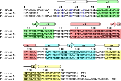
FIGURE 1.
Primary and secondary structure features of FCaBP. Alignment of the primary sequence of T. cruzi FCaBP with that of T. rangeli, and T. brucei. Secondary structural elements indicated schematically were derived from the x-ray structure and analysis of NMR data (3JHNHα, chemical shift index (59), and sequential NOE patterns). The four EF-hands (EF1, EF2, EF3, and EF4) are highlighted in green, salmon, cyan, and yellow, respectively. Residues in the 12-residue Ca2+ binding loops are underlined, and chelating residues are highlighted in bold. Invariant basic residues on the protein surface and implicated in membrane binding are colored blue.

