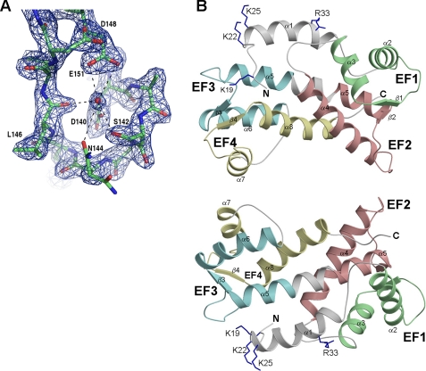
FIGURE 2.
X-ray crystal structure of FCaBP (PDB code 3CS1). A, representative electron density map of FCaBP, showing the Ca2+ binding loop of EF3. A bound water molecule (red sphere) is present in the EF3 Ca2+ binding site of apoFCaBP. B, ribbon diagrams depicting the main chain structure of FCaBP viewed from the membrane binding interface (top) and rotated 180° (bottom). The EF-hands are colored as in Fig. 1.

