
FIGURE 5.

In situ mitochondrial swelling in cortical astrocytes. (A) Raw fluorescence micrographs of a mito-DsRed2-expressing cortical astrocyte, acquired by single-plane wide-field fluorescence microscopy. (B) TR shown as pseudocolor images gated with raw intensity. Solid and open arrows indicate slender and swollen mitochondria, respectively. Open arrowheads indicate swollen, beaded (grain) mitochondria. Scale bar, 5 μm. (C–G) Time course of the TR shown as mean ± SE of single cell traces (gray) normalized to the baseline (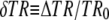 ). (C) Effect of valinomycin (0.2 and 250 nM) and alamethicin (Ala; 40 μg/ml; n = 3). Numbers in black discs refer to the frames shown in A. (D) Switch of superfusion to identical buffer (n = 4). (E) Effect of FCCP (1 μM) and alamethicin (40 μg/ml; n = 3). (F) Effect of antimycin A3 (2 μM) plus oligomycin (2 μM) (n = 4). (G) Effect of mastoparan (10 μM) and alamethicin (40 μg/ml) (n = 3). The filter functions for the TR calculation are given in Fig. 2 B.