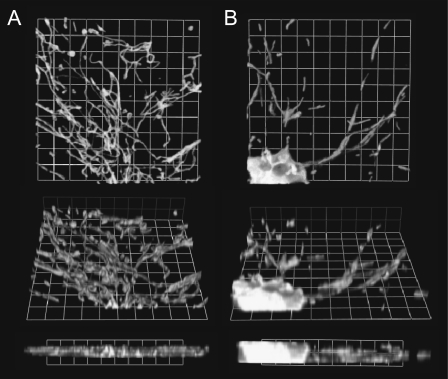
FIGURE 7.
Three-dimensional, volume-rendered reconstruction of confocal image stacks. (A and B) An astrocyte in culture (A) and a cortical neuron expressing mito-DsRed2 (B). Both image stacks were acquired in identical imaging conditions, with the exception that finer z-steps were used for astrocytes. Grid, 3.3 μm. Note that whereas most mitochondria are in a single plane in the astrocyte, mitochondria in neurites are often at different z-coordinates, and only a section of the soma was acquired.

