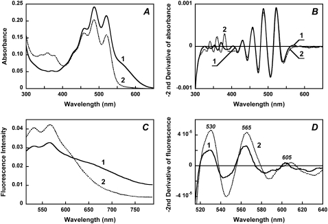
FIGURE 4.
Effect of reduction of the retinal Schiff base with sodium borohydride on the absorption spectrum of xanthorhodopsin and the fluorescence intensity of salinixanthin. (A) Absorption spectra of (1) cell membrane fraction containing xanthorhodopsin, pH 8.5, 100 mM NaCl; (2) after reduction of the retinal Schiff base with sodium borohydride. (B) Second derivatives of the spectra in panel A (multiplied by −1) showing that the sharp carotenoid bands at 521, 486, and 456 nm did not change their shape on retinal Schiff base reduction. (C) Fluorescence spectra: (1) initial, pH 8.5; 2, treated with NaBH4. (D) Second derivatives (multiplied by −1) of the fluorescence spectra of: (1) initial sample; (2) after treatment with NaBH4. The maxima correspond to salinixanthin fluorescence bands.

