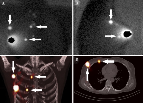
Fig. 2.
Woman with a right breast cancer. The conventional lymphoscintigraphic anterior (A) and lateral (B) images show sentinel nodes in the axilla (descending arrow), in the internal mammary chain (upper horizontal arrow), and a sentinel node that is located in the breast or the internal mammary chain (lower horizontal arrow). The three-dimensional fused SPECT/CT maximum intensity projection (C) depicts the axillary sentinel node (descending arrow), the internal mammary chain node in the second intercostal space (upper horizontal arrow), and shows that the other sentinel node is presumably located in the internal mammary chain (lower horizontal arrow). The transaxial fused SPECT/CT image (D) visualizes the axillary sentinel node (ascending arrow) and confirms that the lower sentinel node is an internal mammary chain node located in the fourth intercostal space (horizontal arrow)

