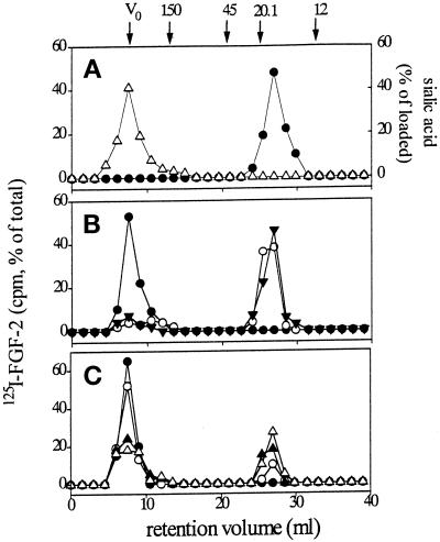
Figure 1.
Size-exclusion chromatography of FGF-2–ganglioside complexes. (A) 100 μl samples containing 3 pmol of 125I-FGF-2 (•) or 125 nmol of GM1 (▵) in PBS were incubated for 10 min at room temperature, loaded separately onto size-exclusion fast protein liquid chromatography Superose-12 column, and eluted in PBS at 1 ml/min flow rate. Radioactivity or NeuAc concentration was measured in each fraction for the evaluation of 125I-FGF-2 or ganglioside content, respectively. (B) 3 pmol of native (•, ○) or of heat-denatured 125I-FGF-2 (▾) were incubated for 10 min at room temperature with 125 nmol of GM1 (closed symbols) or of asialo-GM1 (○). Then, samples were subjected to size-exclusion chromatography as in panel A, and radioactivity was measured in each fraction. (C) 3 pmol of 125I-FGF-2 were incubated for 10 min at room temperature with decreasing concentrations of GM1 [12.5 nmol (•), 5 nmol (○), 1.25 nmol (▴), or 0.125 nmol (▵)]. Then, samples were subjected to size-exclusion chromatography as in panel A, and radioactivity was measured in each fraction. Molecular size standards (in thousands) were ferritin (Mr 440,000), that eluted with the void volume of the column (V0), IgG (Mr 150,000), ovalbumin (Mr 45,000), soybean trypsin inhibitor (Mr 20,100), and cytochrome C (Mr 12,000).

