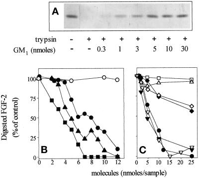
Figure 2.
Protection of FGF-2 from tryptic digestion by gangliosides and related molecules. (A) Representative experiment in which 1 μg aliquots of FGF-2 were incubated at 37° C for 3 h with 60 ng of trypsin in the absence or in the presence of the indicated amounts of GM1. Then samples were analyzed by 15% SDS-PAGE followed by silver staining of the gel. (B and C) Aliquots (1 μg) of FGF-2 were incubated with trypsin in the presence of the indicated amounts of asialo-GM1 (○), GM1 (•), GD1b (▴), GT1b (▪) (panel B) or GM1 (•), NeuAc (□), N-acetylneuramin-lactose (▵), GM2 (▾), GM3 (♦), sulfatide (▿), galactosyl-ceramide (⋄) (panel C). Then samples were analyzed by 15% SDS-PAGE followed by silver staining of the gel. The amount of nondigested FGF-2 was evaluated by soft-laser scanning of the gel, and data are expressed as percentage of digested FGF-2 in respect to samples in which trypsin was omitted. Each point is the mean of two to five determinations in duplicate. SEM never exceeded 13% of the mean value.

