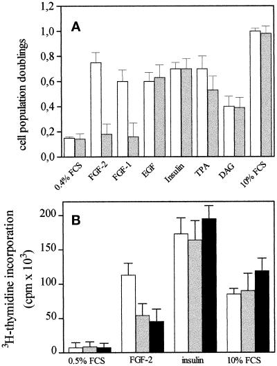
Figure 9.
Effect of GM1 on the mitogenic activity of different endothelial cell mitogens. (A) Subconfluent cultures of GM 7373 cells were incubated for 24 h at 37°C in 0.4% FCS with no addition (control) or with FGF-2 (10 ng/ml), FGF-1 (30 ng/ml), EGF (30 ng/ml), insulin (10 μg/ml), 10% FCS, 12-O-tetradecanoyl phorbol 13-acetate (TPA, 30 ng/ml), or 1,2-dioctanoyl-sn-glycerol (DAG, 5 μg/ml) in the absence (white bars) or in the presence (gray bars) of 10 μM GM1. At the end of incubation, cells were trypsinized and counted in a Burker chamber. Data are expressed as cell population doublings during the 24-h incubation period. Each point is the mean ± SEM of two to six determinations in duplicate. All mitogens induce a statistically significant increase of cell proliferation rate (Student’s t test, p < 0.05). (B) MAE cells were incubated for 2 d with 0.5% FCS. Quiescent cell cultures were then treated with vehicle (0.5% FCS), FGF-2 (30 ng/ml), insulin (10 μg/ml), or 10% FCS in the absence (white bars) or in the presence of 30 μM (gray bars) or 100 μM (black bars) of GT1b. After 16 h, cells were pulse labeled with [3H]thymidine (1 μCi/ml) for 6 h. The amount of radioactivity incorporated into the trichloroacetic acid-precipitable material was measured. Each point is the mean ± SEM of three determinations in triplicate. At both concentrations, GT1b causes a statistically significant decrease of DNA synthesis induced by FGF-2 (Student’s t test, p < 0.05).

