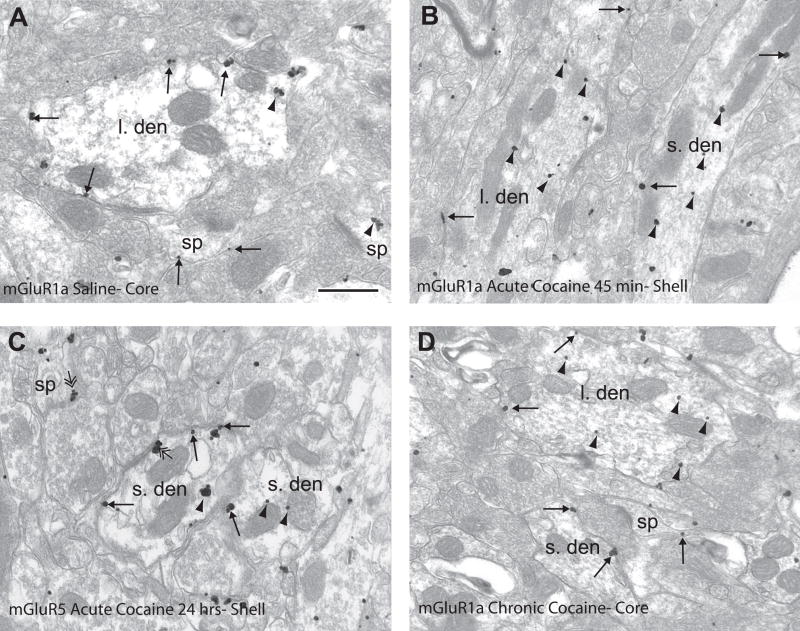
Figure 3.
mGluR1a and mGluR5 immunogold labeling in saline- and cocaine-treated rats. (A) mGluR1a immunogold labeling in the nucleus accumbens core of a saline-treated rat. Note the extrasynaptic (single-headed arrows) labeling on the plasma membrane of the large dendrite (l. den) and spine (sp). (B) mGluR1a immunogold labeling in nucleus accumbens shell 45 minutes following cocaine treatment. Note the increased intracellular labeling (arrowheads) in large and small dendrites (s. den). (C) mGluR5 immunogold labeling in the nucleus accumbens shell 24 hours following cocaine injection. mGluR5 is distributed extrasynaptically (single-headed arrows) and intracellularly (arrowheads) in the dendrites. Also note the perisynaptic labeling to an asymmetric axodendritic and axospinous synapse (double-headed arrow). (D) mGluR1a immunogold labeling in the nucleus accumbens core of a rat chronically treated with cocaine followed by 3 weeks withdrawal. Note the larger amount of intracellular labeling in the large dendrite and extrasynaptic plasma membrane labeling in the small dendrite and spine. Scale bar=0.5μm.

