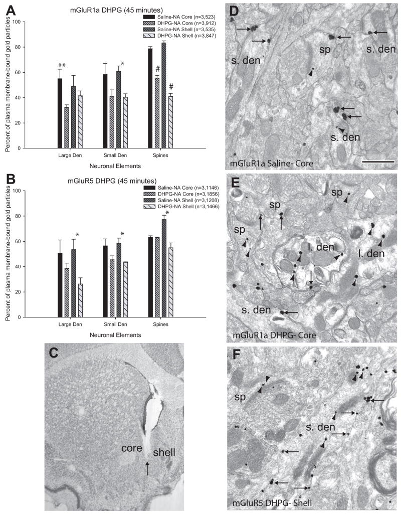
Figure 5.
Histograms and immunogold labeling for mGluR1a and mGluR5 in saline- and DHPG-treated rats. (A) and (B) are summary histograms showing the percentage of plasma membrane-bound labeling for mGluR1a (A) and mGluR5 (B) in the core and shell of the nucleus accumbens of saline- and DHPG-treated rats. Data are presented as mean percentages (±SEM) of gold particles on the plasma membrane of large or small dendrites and spines; in parentheses n= number of animals used, followed by the total number of gold particles counted in each experimental group. One-way ANOVAs and Tukey’s post-hoc tests reveal that there is a lower percentage of plasma membrane-bound mGluR1a in large dendrites of the core (p<0.05, double asterisk), in small dendrites of the shell (p<0.05, single asterisk), and in spines of both shell and core of the accumbens (p<0.001, number signs) in DHPG-treated animals. For mGluR5, there is a significantly lower percentage of labeling on the plasma membrane in the shell of the accumbens on large and small dendrites (p<0.05, single asterisk) and spines (p<0.01, number sign). (C) Light micrograph of a sample DHPG injection into the core and medial shell of the nucleus accumbens. The arrow indicates the tip of the syringe. (D–F) show examples of labeled elements from saline- treated (D) and DHPG-treated animals (E–F). (D) mGluR1a labeling in the core of the accumbens of a saline-treated animal. Note the majority of extrasynaptic labeling (single-headed arrows) on the plasma membrane of small dendrites (s.den) and spines (sp). (E) mGluR1a labeling in the core of the accumbens of a DHPG-treated animal. Notice the increase in intracellular (arrowheads) labeling in both dendrites and spines. (F) mGluR5 labeling in the shell of the accumbens of a DHPG-treated animal. Note the large pool of intracellular labeling in dendrites and spines. Scale bar=0.5μm. Total number of elements examined: mGluR1a: Saline, Core: large den=15, small den=137, spines=72; DHPG, Core: large den=57, small den=170, spines=91; Saline, Shell: large den=24, small den=94, spines=40; DHPG, Shell: large den=50, small den=134, spines=60. mGluR5: Saline core: large den=36, small den=115, spines=111; DHPG, Core: large den=38, small den=190, spines=105; Saline, Shell: large den=34, small den=147, spines=47; DHPG, Shell: large den=32, small den=157, spines=57.

