Figure 6.
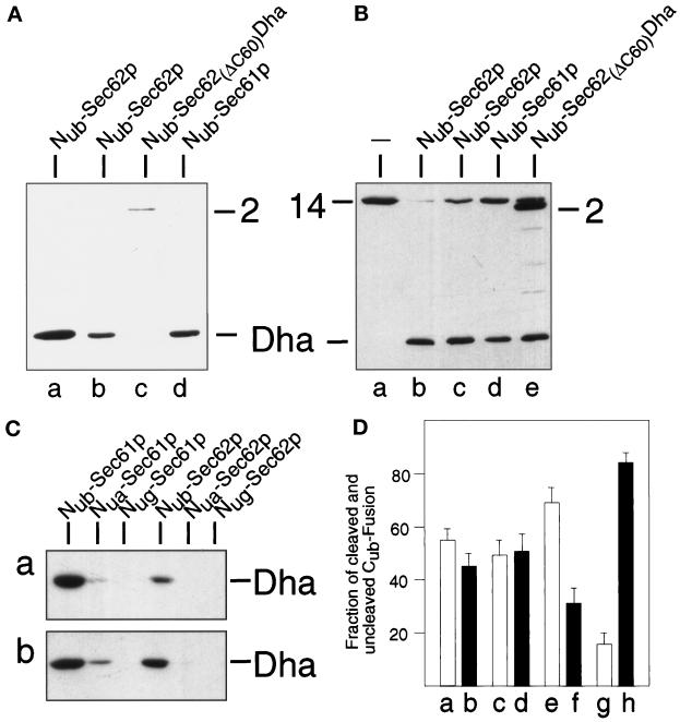
Sec61p, but not a mutant of Sec62p, are close to the nascent chain of a translocated protein. (A) These assays employed S. cerevisiae expressing Mfα37-Cub-Dha (construct 8, Figure 2) and one of the following Nub fusions (Figure 2): Nub-Sec62p, either integrated (lane a) or plasmid borne (lane b); Nub-Sec62(ΔC60)Dha (lane c); and Nub-Sec61p (lane d). Whole-cell extracts from these strains were subjected to immunoblot analysis with anti-ha antibody. (B) Same as panel A but the same Nub fusions were coexpressed with Tpi1-Cub-Dha (construct 14; Figure 2). Numbers 2 and 14 indicate the positions of the corresponding (uncleaved) fusions. (C) Lane a: S. cerevisiae expressing Suc223-Cub-Dha (construct 10; Figure 2) together with either Nub-Sec61p, Nua-Sec61p, Nug-Sec61p, Nub-Sec62p, Nua-Sec62p, or Nug-Sec62p; lane b: same as lane a but cells expressed Mfα37-Cub-Dha (construct 8; Figure 2) instead of Suc223-Cub-Dha. (D) S. cerevisiae cells expressing Mfα37-Cub-Dha together with Nub-Sec61p (a and b) or Nub-Sec62p (e and f), and cells expressing Suc223-Cub-Dha together with Nub-Sec61p (c and d) or Nub-Sec62p (g and h) were labeled for 5 min with 35S-methionine. Whole-cell extracts were immunoprecipitated with anti-ha antibody, followed by SDS-PAGE, and quantitation of the cleaved and uncleaved Cub fusions using PhosphorImager. Shown are the relative amounts of the cleaved (white bars: a, c, e, and g) and uncleaved (black bars: b, d, f, and h) Cub fusions. The sum of a cleaved and uncleaved fusion was set at 100 in each of the three independent experiments. SDs are also indicated.

