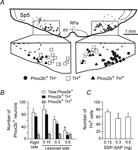
Figure 2. SSP-SAP destroys Phox2b+TH− neurons selectively.
A, computer-assisted plot of the Phox2b+TH− neurons and C1 neurons (TH+) present in a single 3 μm-thick coronal brain section from a rat that had received a single 0.6 ng dose of SSP-SAP on the left side of the brain (Bregma level around −11.6 mm). Note the selective loss of the Phox2b+TH− neurons on the side with the lesion. B and C, group data. Each column represents the total number of neurons of a given type present in 9 consecutive 30 μm-thick coronal sections separated by 180 μm. The middle section was as close as possible to Bregma −11.6 mm. RPa = raphe pallidus, py = pyramidal tract.

