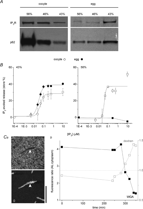
Figure 6. Structural inhibition of IP3Rs in AL.
A, immature oocytes were homogenized and fractionated through a discontinuous sucrose gradient to yield ‘heavy’ vesicles derived from AL (56%), a population of cortical ER vesicles (43%) and an intermediate mixed vesicular population (46%). The same protocol was utilized on naturally shed eggs. Equal amounts of protein from each fraction were subjected to immunoblotting with anti-IP3R or anti-p62 antibodies. B, left, IP3 evoked 45Ca2+ release from the light membrane fraction (43%) extracted from oocytes (○) or eggs (•). Right, IP3 evoked 45Ca2+ release from the heaviest membrane fraction (AL) extracted from oocytes (○) or eggs (•). C, altered membrane architecture of AL following NPC dissociation: a, image stills showing distribution of a 70 kDa fluorescent dextran (top) and a fluorescently tagged WGA conjugate (bottom) with respect to the same AL (white arrows) before maturation (t= 0 min, scale bar = 20 μm); b, ratio of fluorescence of WGA (▪, left ordinate) and dextran fluorescence (□, right ordinate) labelling of AL relative to randomly chosen surrounding cytoplasmic area at the indicated time points during maturation (minutes). Values below dashed grey line (ratio = 1, right ordinate) indicates exclusion from AL.

