
Fig. 6.
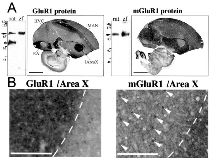
Differential expression at the protein level. A: Right: Western blots of rat and zebra finch (zf) whole-brain extracts reacted with antibodies that recognize rat GluR1 and mGluR1 carboxy termini. Cross-reactivity to similarly sized zebra finch proteins are found (arrows). *Additional protein band detected in rat but not in zebra finch brains. It is unknown whether this additional band is a specific variant of rat GluR1. Left: Immunohistochemistry for GluR1 and mGluR1 protein expression in sagittal zf male brain sections with the same antibodies. B: Higher magnification showing protein expression of GluR1 and mGluR1 in l-Area X (left of the dashed lines) and surrounding striatum. Arrowheads point to the sparse, large neurons in Area X. The clear white areas seen in the mGluR1 image of B are blood vessels that have expanded upon perfusion of the animal. Scale bars = 2 mm in A; 300 μm in B.