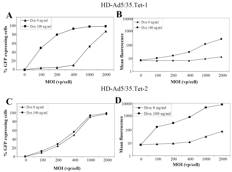
Fig. 3. Transduction studies in Mo7e cells.
MO7e cells were infected with HD-Ad5/35.Tet-1 (A,B) or HD-Ad5/35.Tet-2 (C,D) at increasing MOIs of 100, 200, 400, 1000, 2000 vp/cell. One set of infected cells was left non-induced, to the other set of cells, 100ng/ml Doxycyclin were added for induction. Twenty-four hours after infection/induction, the percentage of GFP expressing cells (A,C) and mean GFP fluorescence (B,D) were measured by flow cytometry.
N=3. The SEM was consistently less than 10% of the Mean.

