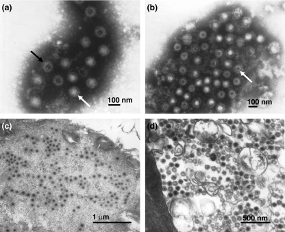
Figure 1.
(a) Negative staining of Minaçu virus, in fluid of VERO cells (5 days p.i.), showing complete (white arrow) and incomplete (black arrow) viral particles (114,000×). (b) Electronic immunomicroscopy of Minaçu virus and homologous serum showing viral particles (arrow) surrounded by a dense antibody halo at the dilution 1:500 (90,000×). (c, d) Electronic micrographs of Minaçu virus in ultra-thin sections of VERO cells (4 days p.i.). Note the viral particles within the cell cytoplasm (60,000×) (c) and the absence of a viral envelope (80,000×) (d).

