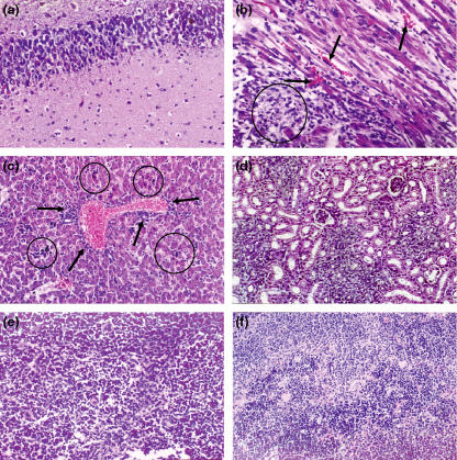
Figure 3.
Histological changes in the brain, heart, liver and kidneys of suckling mice intracerebrally infected with the Minaçu virus, 6 days post-infection. (a) Brain with extensive area of lithic necrosis −200×; (b) heart with necrotic areas (circle), micro-haemorrhages (arrows) and intense oedema −400×; (c) liver with focal areas of cellular necrosis, and acidophilic corpuscles (circles). Note the perivascular inflammatory infiltrate in the portal space (arrows) −200×; (d) kidney with slightly tumefied renal tubules and slightly congested interstitial area −200×; (e) spleen showing cellular necrosis in red pulp – 200×; (f) spleen with cellular necrosis and haematic infiltration – 200×.

