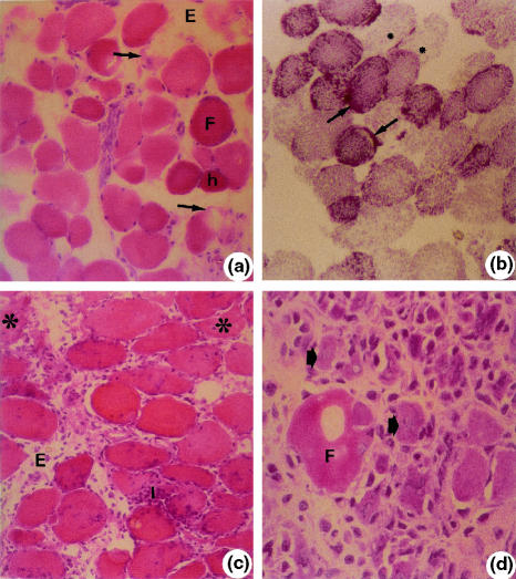
Figure 2.
Soleus muscle. a (IR-1 h). Round (F). Hypercontracted fibres (h). Fibre with myofibril disorganization (arrows). Oedema (E). Haematoxylin–eosin (HE) 40×; b (IR-1 h). Round fibres with irregular distribution of oxidative metabolism enzymes (granulations) (arrows). Lack of enzyme activity (*). NADH-TR 40×; c (IR-24 h). Intense inflammatory infiltrate (I). Oedema (E). Necrotic fibres (*). HE, 40×, d (IR-72 h). Necrotic fibre (F). Small basophilic fibres with central nucleus (arrows). HE, 100×.

