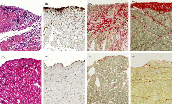
Figure 2.
Photomicrographs depicting non-infarcted subendocardial regions of the left ventricle in the rat model of infarction. Top panels represent rats with myocardial infarction and bottom panels sham animals. Left panels show inflammatory cell reaction in HE-stained tissue sections at day 3. Left middle panels show myofibroblasts positive to α-smooth muscle actin immunostaining (brown cytoplasm) in tissue sections co-stained with haematoxylin at day 7. The last two right panels show red stained fibrosis in Sirius red-stained tissue sections at days 7 and 28 respectively. Prominent differences in each of these phases of subendocardial remodelling of the non-infarcted left ventricular myocardium can be observed between infarcted and sham rats.

