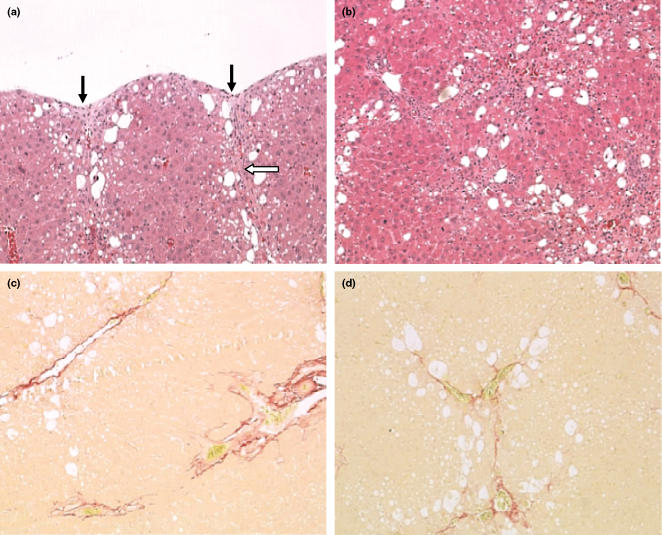
Figure 2.
Histopathology of the liver in rats treated with carbon tetrachloride (CCl4) twice a week for 6 weeks and sampled at that time. (a) Liver from a rat treated with CCl4 at 0.6 ml/kg twice weekly for 6 weeks. In the subcapsular region, there is hepatocellular vacuolation, degeneration and necrosis, with capsular pitting or contraction (black arrows). There is collagen formation in the capsular area which extends downwards into the liver parenchyma (open arrow). Original magnification of image; ×100 [haematoxylin and eosin (H&E)]. (b) Liver from a rat treated with CCl4 twice weekly for 6 weeks at 1.0 ml/kg. There is significant fibrosis which is tending to link centrilobular and periportal areas. Hepatocytes are vacuolated with evidence of degeneration. Original magnification of image; ×100 (H&E). (c) Liver from a rat treated with CCl4 twice weekly for 6 weeks at 0.7 ml/kg; there is perivascular fibrosis in the centrilobular and periportal areas, with some evidence of bridging along the sinusoidal tracts. Original magnification of image; ×100 (Sirius red). (d) Liver from a rat treated for 6 weeks, twice weekly at 1.0 ml/kg CCl4. Perivascular fibrosis is evident with bridging along sinusoidal tracts within the liver parenchyma. Original magnification of image; ×100 (Sirius red).

