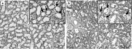
Figure 1.
Conventional haematoxylin-and-eosin-staining of the control lachrymal gland (a) shows the typical histological tubulo-acinar structure of the inferior lachrymal gland and the cubic regular shape of acinar cells with basally located nuclei (arrows). Three days after irradiation with 15 Gy, vacuolopathy and secretory retention were evident in the end-piece epithelium (b; arrows) while ductal cells stayed unaffected.

