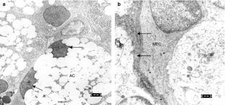
Figure 3.
(a) Electron microscopic appearance of the lachrymal acinar cells (AC) 72 h after irradiation showing granula retention and peripheral displacement of the cell nuclei (arrows). Bar = 2.5 µm. (b) Thickening of the basal membrane (arrows) as well as condensation of the myofilaments in the myoepithelial cells (MECs) noticed 72 h after irradiation (asterisks). Bar = 0.6 µm.

