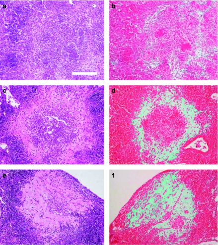
Figure 4.
Evolution of intragranulomatous necrosis in pulmonary granulomas (INPG) through time in mice C57B/6 infected by the Mycobacterium tuberculosis strain UTE 0335R at week 3 (a, b), 6 (c, d) and 9 (c, f) post-infection (Experiment C). Panels (a), (c), (e) represent cuts stained with haematoxylin and eosin (H&E). Panels (b), (d), (f) represent cuts stained with Trichromic of Masson which reveals the progression from the periphery to the centre of the collagen-based fibrotic tissue (in green) to occupy the necrotic centre of a primary granuloma. Haematoxylin and eosin stain shows a mixture of monocytes and neutrophils at week 3; a centre occupied by debris, karyorrhexis, fibrin and some neutrophils at week 6 and a compact eosinophilic scar at week 9. Bar represents 100 µm.

