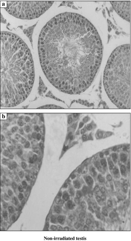
Figure 1.
Histological features of the non-irradiated testis. The seminiferous tubule is formed of the lining germinal epithelium (spermatogonia Type A and Type B, primary spermatocytes and spermatids) and the supporting Sertoli cells. Aggregates of interstitial cells are present in between tubules (a, ×200 and b, ×1000).

