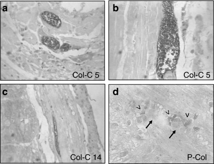
Figure 4.
Immuno-histochemical labelling of Trypanosoma cruzi amastigotes by peroxidase: (a) Section of the heart with mononuclear cells infiltration and the presence of intracellular amastigote forms of T. cruzi. (×400). (b) Skeletal muscle showing a large nest of amastigote forms immunolabelled with anti-T. cruzi antibodies. (×400). (c) Clone Col-C5 – section of the intestine, showing amastigotes of T. cruzi inside the smooth muscle cells of the intestinal wall. (×100). (d) Clone P-Col (parental strain) – presence of amastigotes (arrow head) in the Auerbach plexus (arrows) of the intestinal wall, ×400.

