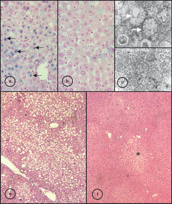
Figure 2.
Liver sections of 18-month-old β2m and Hfe-/mice. Sections stained for iron by Perls method, the blue stain indicates heavy iron deposition: (a) β2m-/- strain, black arrow shows blue stained hepatocyte nuclear inclusions; (b) Hfe-/- mice (original magnification ×400). Electron photomicrograph of hepatic parenchyma cells; (c) β2m-/- mice, white arrow indicate abnormal mitochondria without visible cristae; (d) Hfe-/- strain (original magnification ×8000). Liver sections stained with HE; (e) β2m-/- mice, large vacuoles visible trough the parenchyma indicative of generalized steatosis; (f) Hfe-/- strain, asterisk indicate steatosis foci (original magnification ×40).

