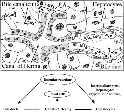
Figure 9.
Schematic representation of normal liver structure and changes of its elements during disease and regeneration. (a) The lobular structure with the canal of Hering which drains bile from the bile canaliculi into the bile duct [modified from L.C. Junqueira & J. Carneiro (1980). In Basic Histology, Lange Medical Publications, p. 350]. (b) Oval cells can proliferate from the canals of Hering and lead to ductular epithelial structures. These proliferating cells are regarded as progenitor cells or intrahepatic stem cells which can differentiate via intermediate-sized hepatocytes into mature hepatocytes and bile duct epithelium; also, extra-hepatic stem cells of bone marrow origin are discussed to be a source of oval cells and provide progenitor cells for liver regeneration.

