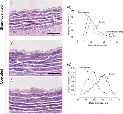
Figure 3.
High resolution light microscopy. Representative views of the prestenotic segments of the aorta from operated rats (b and c) and the corresponding segments from controls (a). In the prestenotic segment, there is diffuse intimal thickening with enlarged endothelial cells (b) and diffusely distributed minute foci of intimal thickening composed of smooth muscle cells and occasional mononuclear cells with collagen and elastic fibres surrounding then (c) and medial thickening (b and c), contrasting with the delicate structure of the intima in control group (a). The percentile frequency distribution of intimal thickness in the prestenotic segment demonstrates a shift to the right of the values in comparison with the values in corresponding segment in sham-operated animals. Moreover, the numerous discrete foci of intimal thickening, absent in the control aortas, can be clearly demonstrated (d). The same can be seen when the percentile frequency distribution of media thickness is plotted clearly, demonstrating the medial thickening in operated animals in comparison with controls (e). Scale bars, 20 μm (a–c).

