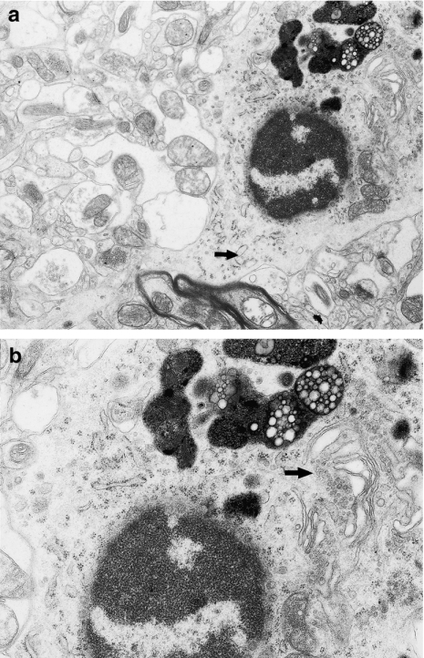
Figure 4.
(a) Phagocytic microglial cell in a close vicinity of swollen neuropil elements. The nucleus of this cell is round and contains dense heterochromatin, highly clumped under nuclear envelope and sparse euchromatin; in the swollen cytoplasm visible profiles of granular endoplasmic reticulum (→). Neocortex. Nine months of valproate administration. Original magnification ×4000. (b) Higher magnification (×12,000) of phagocytic microglial cell. Well-developed frothy lipophagosomes and lipofuscin-like bodies and dilated Golgi apparatus (→).

