
Fig. 4.
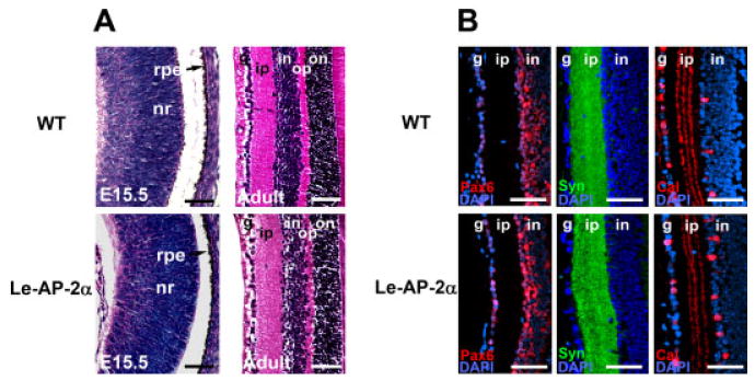
Retinogenesis in Le-AP-2α mutant mice. A: At embryonic day (E15.5), analysis of wild-type (top panels) and Le-AP-2α (bottom panels) embryos indicates no significant change in retinal development, while examination of adult eyes indicates no change in retinal lamination between control and Le-AP-2α mice. B: Immunofluorescent staining of paraffin-embedded sections for the retinal cell markers, Pax6 (red), syntaxin-1 (green), and calretinin (red) revealed no significant alteration in protein expression within the adult retinas of Le-AP-2α mutant mice compared to control littermates. Nuclei are counterstained with 4′,6-diamidine-2-phenylidole-dihydrochloride (DAPI, blue). nr, neural retina; rpe, retinal pigmented epithelium; g, ganglion cell layer; ip, inner plexiform layer; in, inner nuclear layer; op, outer plexiform layer; on, outer nuclear layer. Scale bars = 100 μm.