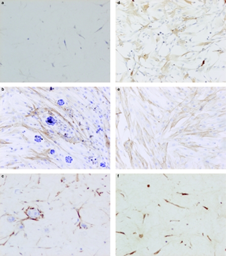
Figure 3.
Micrographs showing α-smooth muscle actin (α-SMA) expression in cultured and cocultured tumour and stromal cells. (a) Stromal cells cultured in gels in medium with 1% fetal calf serum; less than 5% of the cells are positive for α-SMA (ABC, haematoxylin counter-stain ×200). (b) Coculture of stromal cells with HT-29 cells on monolayers; more than 90% of the cells expressing α-SMA (ABC, haematoxylin counter-stain ×200). (c) Coculture of stromal cells with Caco-2 in gels; more than 90% of the cells expressing α-SMA (ABC, haematoxylin counter-stain ×200). (d) Stromal cells cultured on monolayers with HT-29-conditioned media in monolayers. Increase in the number of stromal cells expressing α-SMA (ABC, haematoxylin counter-stain ×200). (e) Stromal cells cultured on monolayers, addition of TGF-β; more than 90% of the cells expressing α-SMA (ABC, haematoxylin counter-stain ×200). (f) Stromal cells cultured in gels, addition of TGF-β; more than 90% of the cells expressing α-SMA (ABC, haematoxylin counter-stain ×200).

