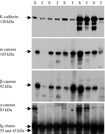
Figure 4.
Immunoprecipitation of the E-cadherin/catenin complex in the TX-100-soluble (S) and TX-100-insoluble (I) cell protein fractions, using the anti-E-cadherin antibody. Bands of α-, β- and γ-catenin are seen in both fractions of all cell lines; SW480 (lanes 1 and 2), SW620 (lanes 3 and 4), HCT116 (lanes 5 and 6), HT29 (lanes 7 and 8) and Caco-2 (lanes 9 and 10).

