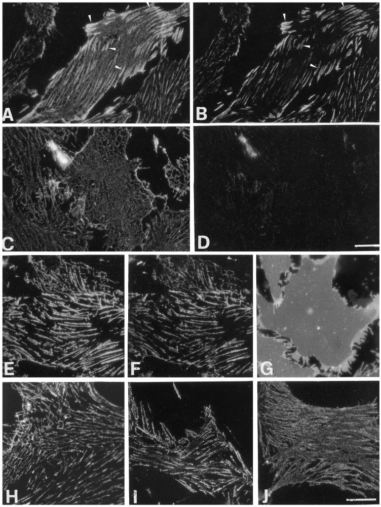
Figure 1.
.Calcium-dependent redistribution of β1 integrins in VPMs. VPMs from adherent CEFs were fixed after preparation on ice (A and B) or after incubation for 15 min at 37°C in HCB (C and D), in LCB (E–G), in HCB followed by 15 min at 37°C in LCB (H), or in LCB followed by 15 min at 37°C in HCB (I). (J) VPMs were treated as in I, except that 200 μg/ml integrin β1 function-blocking antibody CSAT was present during the last 15 min incubation at 37°C in HCB. For staining with the anti-integrin β1 TASC mAb (B, D, and F), VPMs were incubated with a 20 μg/ml concentration of this antibody for 20 min at 0°C before fixation. VPMs were then fixed and processed for immunofluorescence. (A, C, E, and H–J) staining with the β1-cyto polyclonal antibody. Primary antibodies were revealed by FITC-conjugated anti-rabbit IgG and TRITC-conjugated anti-mouse IgG, respectively. (G) VPMs were stained with the lipophilic carbocyanine dye DiIC16. The same fields are shown in A and B, in C and D, and in E and F. Arrowheads in A and B point to focal adhesions, where colocalization of β1-cyto and TASC staining is visible. Bars, 10 μm.

