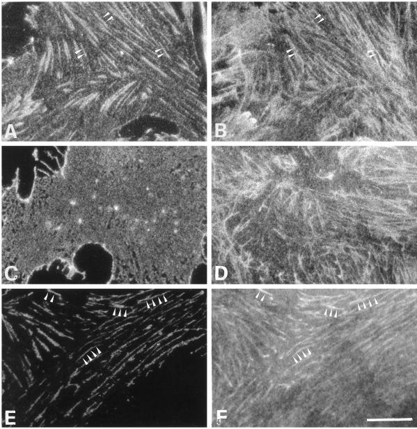
Figure 2.
Localization of β1 integrins and ECM fibrils after treatment of VPMs at different calcium concentrations. VPMs from CEFs were fixed immediately after preparation on ice (A and B), after incubation for 15 min at 37°C in HCB (C and D), or after incubation for 15 min at 37°C in LCB (E and F). Fixed VPMs were processed for immunofluorescence using the β1-cyto antibody (A, C, and E) and the M2D5 mAb recognizing fibronectin (B, D, and F). Primary antibodies were revealed by FITC-conjugated anti-rabbit IgG and TRITC-conjugated anti-mouse IgG. The same fields are shown in A and B, C and D, and E and F. Arrowheads in A, B, E, and F point to sites where colocalization of integrins with ECM fibrils is visible. Bar, 10 μm.

