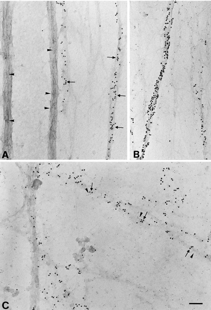
Figure 3.
Ultrastructural localization of β1 integrins along ECM fibrils at low [Ca2+]. VPMs were incubated for 15 min at 37°C in LCB and processed for immunoelectron microscopy as described in MATERIALS AND METHODS. (A) Double labeling with the β1-cyto antibody (large, 15-nm gold; arrows) and the X1E8 mAb decorating actin stress fibers (small, 6-nm gold; arrowheads). (B) Double labeling with the anti-β1 TASC mAb (small, 6-nm gold) and the AAL20 polyclonal antibody against actin, decorating actin stress fibers (large, 15-nm gold). (C) Fibrillar structures are labeled both by the β1-cyto antibody (large gold particles) and by the mAb M2D5 recognizing ECM (small gold particles). In C, a few examples are shown of colocalization of β1 integrins (arrows), with ECM (arrowheads) along fibrils. Bar, 200 nm.

