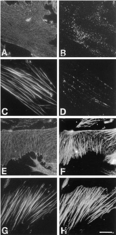
Figure 8.
Reconstitution of the binding of α-actinin to VPMs in the presence of actin stress fibers. After a first incubation for 10 min in low-ionic-strength, high-[Ca2+]–containing buffer, VPMs were incubated for a further 10 min in the same buffer in the absence (A–D) or presence (E–H) of purified α-actinin, as described in MATERIALS AND METHODS. After fixation, F-actin was detected with FITC-phalloidin (C and G), β1 integrins were detected with the β1-cyto antibody (A and E), and α-actinin was detected with a specific mAb (B, D, F, and H). Same fields are shown in A and B, C and D, E and F, and G and H. Bar, 10 μm.

