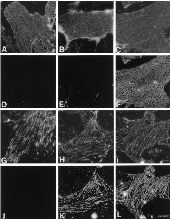
Figure 9.
Reconstitution of the binding of α-actinin to VPMs in the absence of F-actin. VPMs were incubated for 10 min in low-ionic-strength buffer containing 4 μM gelsolin at high [Ca2+] (A–F, I, and L), or low [Ca2+] (G, H, J, and K). The samples were further incubated at 37°C in a buffer with high [Ca2+] (A–F, I, and L), or low [Ca2+] (G, H, J, and K). (C, F, H, I, K, and L) The second incubation was in the presence of purified α-actinin, as detailed in MATERIALS AND METHODS. Membranes were then washed and fixed for immunofluorescence. For low [Ca2+]-induced α-actinin redistribution (I and L), after the second incubation at 37°C for 10 min with α-actinin in high [Ca2+]-containing buffer, the samples were immediately transferred to low [Ca2+]-containing buffer and incubated for 10 min at 37°C to induce integrin redistribution along ECM fibrils before fixation. (A–C and G–I) Integrin distribution, by using the β1-cyto polyclonal antibody. (E, F, and J–L) Localization of α-actinin with the specific mAb. (D) F-actin localization with TRITC-phalloidin. Same fields are shown in A and D, B and E, C and F, G and J, H and K, and I and L. Bar, 10 μm.

