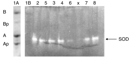
Figure 3.
Basic urea-polyacrylamide gel electrophoresis of spinal cord extract. Except for lane 1 A SOD activity was detected by disappearance of blue staining. Lane (1) bovine α-crystallin, containing the non-phosphorylated (αA and αB) and mono-phosphorylated α-crystallins (αAp and αBp): lane 1A stained with amido black, showing the positions of the α-crystallin subunits on a urea-gel run in parallel; lane (1B) bovine α-crystallin corresponding to lane 1 A; lane (2) transgenic mouse F7 1 female; lane (3) control mouse F7 2 female; lane (4) control mouse F7 3 female; lane (5) transgenic mouse F7 17 male; lane (6) transgenic mouse F7 18 male; lane (7) transgenic mouse F7 98 male; lane (8) transgenic mouse F7 99 male. X = blank.

