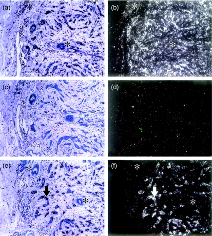
Figure 1.
In situ hybridization. Panels a and b, β-actin-positive control showing expression in both benign and malignant epithelium; panels c and d, negative control (of probe RHK3) negative for both tumour and benign compartments; panels e and f, 11AT1 riboprobe showing abundant expression in the invasive tumour epithelium (arrow) but not in the benign epithelium (asterisks). Panels a, c and e, under conventional light microscopy; b, d and f, under dark field reflected light microscopy.

