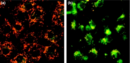
Figure 4.
Representative photographs of rat gastric mucosal (RGM-1) cells after treatment with NOC-18 by JC-1-dye staining. Synchronized images of green (538 nm) and red (590 nm) fluorescence of JC-1-dye staining;(a) the untreated cells and (b) the cells treated with NOC-18. Granular accumulations of the JC-1 dye, emitting red fluorescence in mitochondria, were detected in the untreated cells, but the dye in the cells treated with NOC-18 was diffusely extended in the cytosol, emitting green fluorescence, indicating mitochondrial depolarization in the latter.

