
Figure 3. Identification of Candidate Akt Substrates in Intact Mesangial Cells.
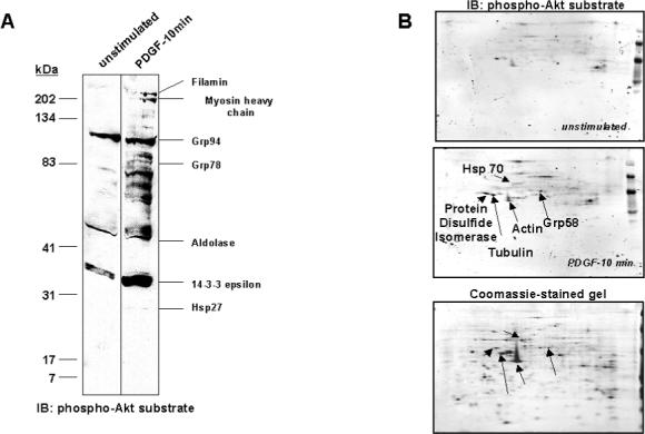
Mesangial cells were cultured in 0.5% FBS/DMEM for 24 hr, prior to incubation with 10 ng/ml PDGF-BB for 10 min, or were left untreated. Lysates were prepared in urea/thiourea buffer and proteins separated by SDS-PAGE (A) or 2-DE (B), on duplicate gels. One of each duplicate gel was stained with Coomassie blue. Proteins from the other gel were transferred to nitrocellulose and immunoblotted with anti-phospho-Akt substrate antibody. Candidate Akt substrates were identified by excision of protein spots/bands from gels, which corresponded to proteins appearing on respective immunoblots, followed by trypsin digestion and identification using peptide mass fingerprinting. Candidate substrates identified are labeled on each figure.