


Figure 7. Akt associates with the microsomal fraction in mesangial cells.
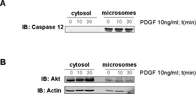
Unstimulated and 10 and 30 min PDGF-stimulated mesangial cells were fractionated into cytosolic and ER-containing microsomal fractions, and the presence of Akt in the two compartments was determined by immunoblot analysis. Panel A is an immunoblot showing for caspase 12 confirming cytosolic and microsomal separation. Procaspase 12, ∼ 55 kDa, is only present in the microsomal fraction. Panel B is an immunoblot of cytosolic and microsomal fractions for Akt showing that Akt is present in both the cytosolic and microsomal fractions and the amount of Akt does not change with PDGF stimulation.