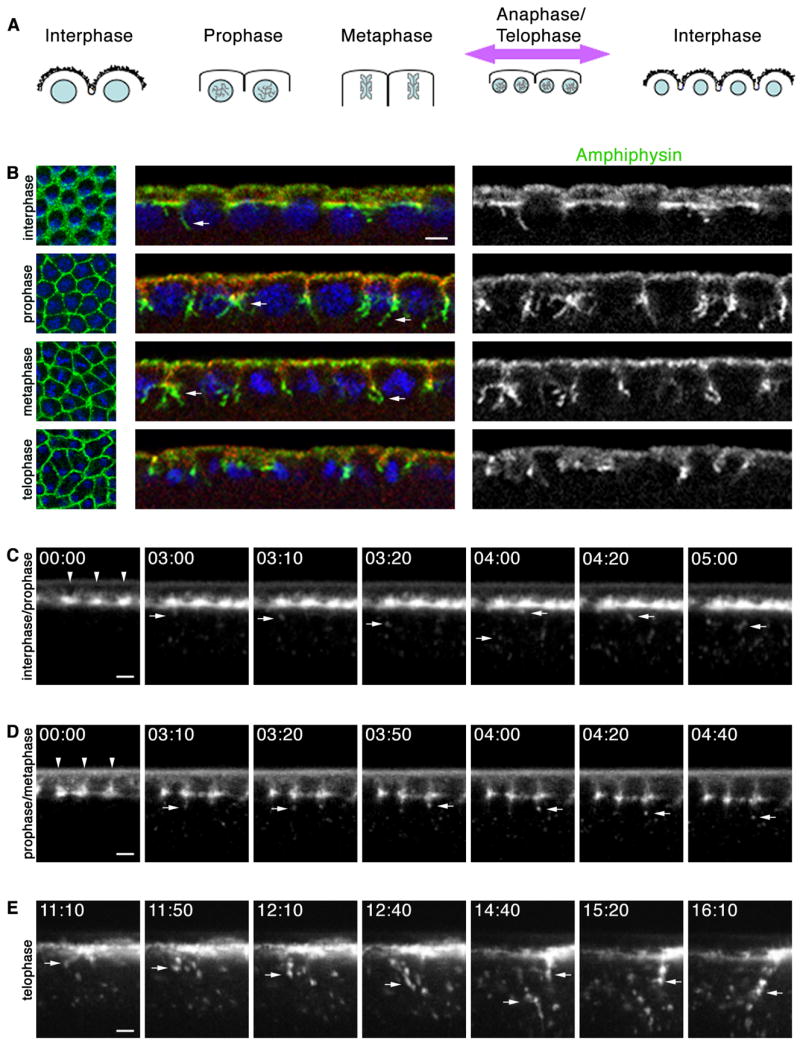
Figure 1.

Cell-cycle progression regulates endocytosis at early mitotic cycles. (A) Furrow dynamics at mitotic cycles (DNA, blue; somatic buds, jagged black lines). Cortex is maximally displaced when metaphase furrows regress (purple arrow). (B) Cross-sections at early mitotic cycle show few Amph (green) tubules (arrows) extend from somatic bud margins (Septin; red) at interphase, many tubules extend from metaphase furrow tips at prophase-metaphase, and no tubules extend from regressing metaphase furrows at telophase. DNA (blue) shows phase of cell cycle. (C–E) Time-lapse cross-sections after peri-vitelline injection of Alexa488-WGA. 00:00 time point set relative to start image acquisition (min:sec). (C) When injected at interphase, Alexa-488-WGA concentrates at somatic bud margins (arrowheads). As embryo enters prophase, vesicles are released from margins (arrows). (D) When injected at interphase/prophase transition, Alexa488-WGA labels ingressing furrows (arrowheads). Vesicles (arrows) are released from furrow tips. (E) When injected at interphase, Alexa488-WGA patches blur as mitosis progresses. First frame shows onset of cortical displacement (large arrow) at late anaphase/telophase when metaphase furrows regress and dramatic endocytosis ensues (small arrows). See Movies S2, S3, S4. Bars are 5 μm.
