
Figure 2.
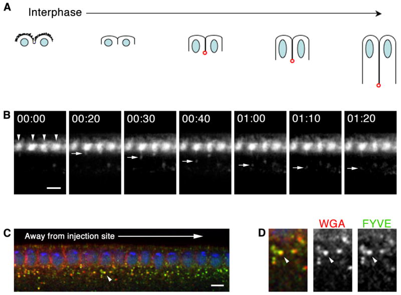
Local endocytosis occurs at forming cellularization furrows. (A) Furrow dynamics at cellularization (DNA, blue; somatic buds, jagged black lines; furrow canals, red). (B–D) Cross-sections after peri-vitelline injection of Alexa488-WGA. (B) Time-lapse shows Alexa488-WGA concentrates at somatic bud margins (arrowheads). Vesicles bud as cellularization furrows form. 00:00 time point set relative to vesicle budding (min:sec). See Movie S2. (C) Peri-vitelline injection of Alexa546-WGA (red), followed by 15 minute chase and fixation. Embryo expresses FYVE-GFP (green). WGA binds embryo surface immediate to injection site. WGA label lengthens basally between nuclei (DNA; blue) as furrows ingress. WGA vesicles incorporate into early endosomes (arrowhead). (D) Higher magnification view of early endosome indicated in B by arrowhead shows Alexa546-WGA (red) and FYVE-GFP (green) co-localize. Bars are 5 μm.