
Figure 6.
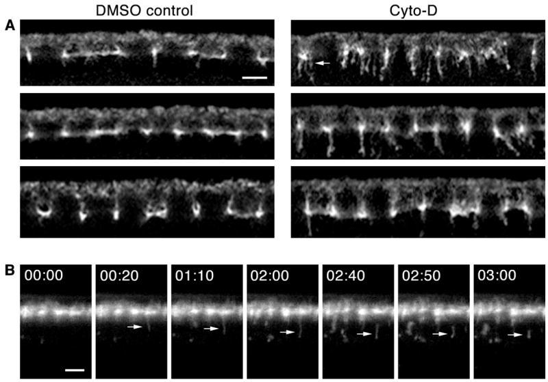
F-actin disruption impairs endocytic scission at cellularization. (A) Cross-sections of Cyto-D versus DMSO control embryos. Amph tubules increase in number after Cyto-D treatment. At earliest cellularization multiple tubules extend from furrow tips (arrow). (B) Time-lapse cross-sections after co-peri-vitelline injection of Alexa488-WGA and Cyto-D. Some budding vesicles distend into tubules (arrow) that persist several minutes before release. 00:00 time point set relative to vesicle budding (min:sec). See Movie S4. Bars are 5 μm.