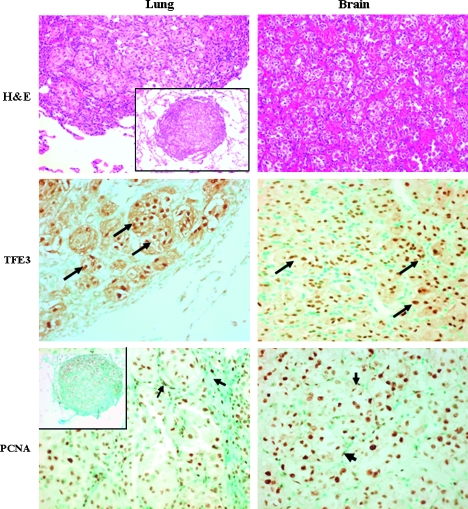
Figure 1.
Pulmonary and brain ASPS metastases. Hematoxylin and eosin staining shows an organoid, pseudoalveolar large round to polyhedral eosinophilic cells displaying large nucleus with a uniform “small nest” pattern separated by fine fibrous septa (original magnification, x100; insert's original magnification, x40). The pulmonary and brain tumor cells express high levels of TFE3 (arrows; original magnification, x200). Both the metastatic cells and the stroma cells (arrows) stain positive for proliferating cell nuclear antigen (original magnification, x200) suggesting that they are at their proliferative state.

