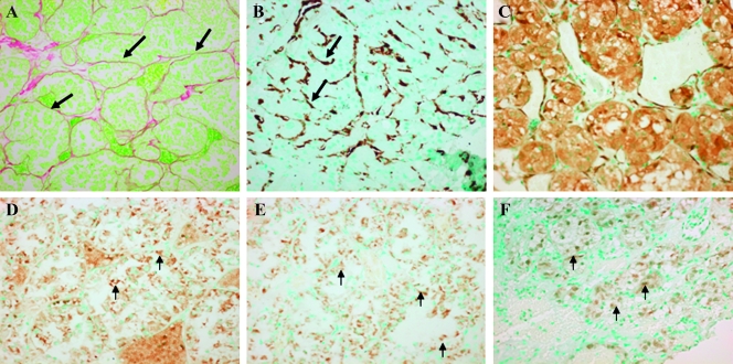
Figure 5.
Stroma components and Smad signaling in the ASPS brain metastases. The tumor cells in the brain metastases are surrounded by fibrous septa (A; original magnification, x100) and populated by myofibroblasts displaying a large amount of αSMA (B; original magnification, x100). The tumor cells exhibited high levels of cytg/STAP (C; original magnification, x100), SRF (D; original magnification, x400) Smad3 (E; original magnification, x400), and phospho-Smad3 (F; original magnification, x100; insert's original magnification, x400).

