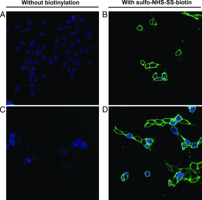
Figure 1.
Biotinylation of plasma membranes using sulfo-NHS-SS-biotin. MDA-MB-231 and MDA-MB-231-B02 were left unlabeled (A and C, respectively), or biotinylated using sulfo-NHS-SS-biotin (B and D, respectively). Biotin labeling was revealed using streptavidin-Alexa 488. Nuclei were stained with TO-PRO-3 fluorescent dye. Confocal images were generated using a Leica TCS SP at an original magnification of x630. Data shown are representative pictures of repeated experiments.

